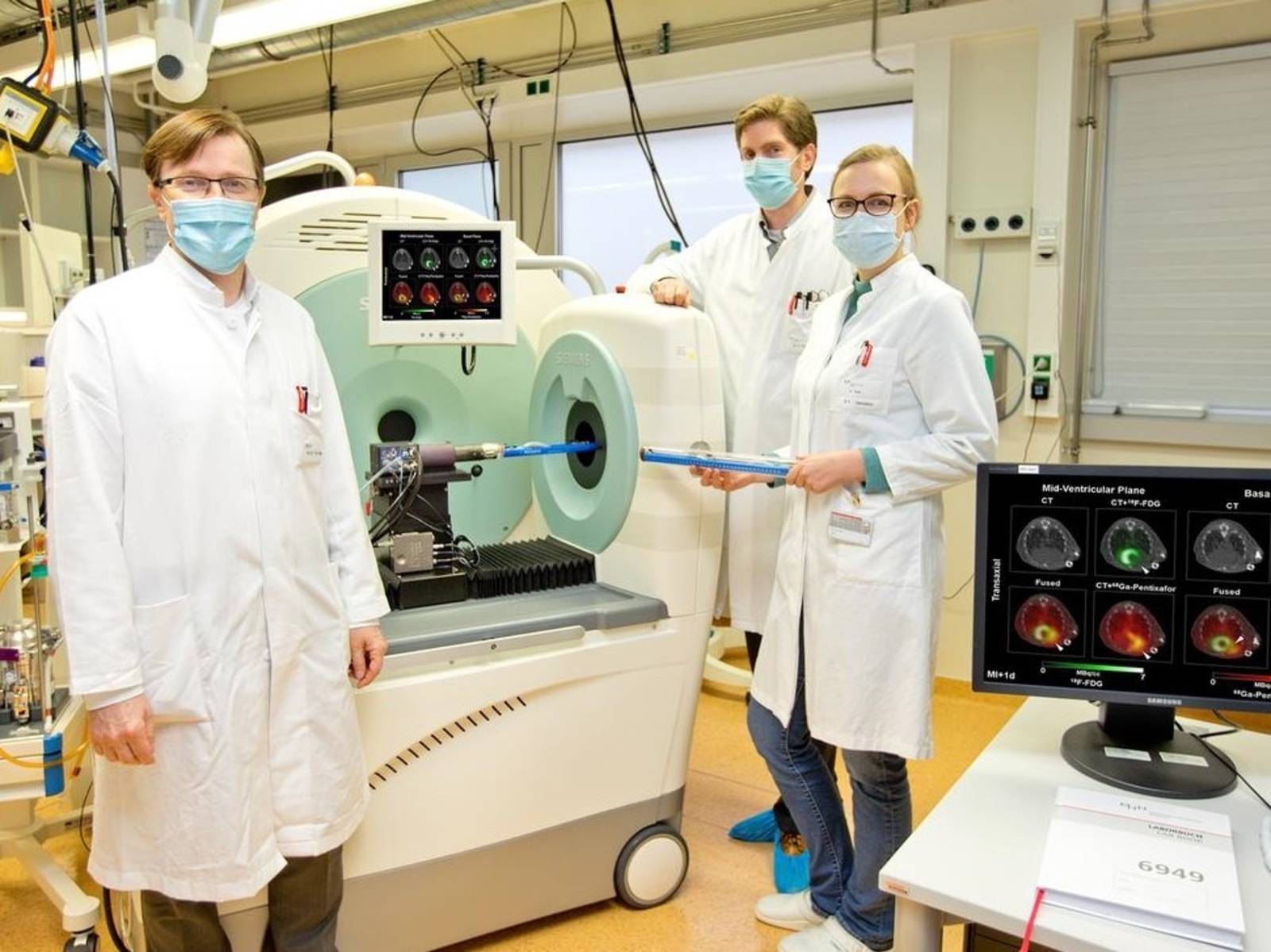
Every year, some 220,000 people in Germany suffer a heart attack (myocardial infarction). This occurs when a blood vessel in the cardiac muscle is blocked, resulting in the heart no longer receiving sufficient oxygen, so that an area of the cardiac muscle dies. Specialized white blood cells (leucocytes), part of the immune system, then cause an inflammatory reaction in the cardiac muscle; this involves breakdown of damaged tissue, thus initiating the healing process. If this inflammatory response is too strong, there is an increased risk of patients suffering from chronic heart failure (cardiac insufficiency). A team of researchers led by Professor Frank Bengel, director of the Department of Nuclear Medicine at Hannover Medical School (MHH), has now found a way of not only accurately monitoring the body’s own repair of the cardiac muscle, but also of improving this repair process. This involves a high-resolution molecular-imaging technique. The study, headed up by James Thackeray, Ph.D., in collaboration with MHH’s Department of Cardiology, has been published in the European Heart Journal, a renowned periodical.
Post-heart attack processes revealed
Using radioactive materials called radiotracers, the research team found out precisely which processes take place following a heart attack. These minute tracing substances, which are mildly radioactive for a brief period, can be made visible in a high-resolution positron emission tomography (PET) scan. The scientists’ focus was on specific proteins in the surface membrane of cardiac-muscle cells. These receptors, named CXCR4, are the binding sites for small signalling proteins (chemokines) that trigger leucocyte migration. "In our investigations, we were able to show that the CXCR4 chemokine receptor may be temporarily upregulated after cardiac-muscle infarction,” explains Annika Hess, Ph.D., the study’s lead author. "This increases the risk of adverse disease progression and development of cardiac insufficiency.”
Treatment tested in investigations with mice and humans
Especially for this study, the investigators produced a radiotracer that was developed in collaboration with the Technical University of Munich (TUM). Once injected into the body, this substance specifically adheres to the CXCR-4 binding site of the white blood cells in the cardiac muscle. PET scanning allows the inflammatory reaction in the heart to be directly visualized without the need for any further intervention. A further advantage of such non-invasive imaging is that this tracer method does not influence the response in the body, hence not leading to false results. In another experiment, the team of researchers was also able to demonstrate how post-infarction healing can be improved and the risk of cardiac insufficiency decreased. “We used a drug that binds at the same site as the tracer, thus blocking the CXCR4 receptor,” Bengel explains. As part of the study, this treatment option was explored by the scientists using a mouse model. They observed that the time of drug administration also appears to be a key factor: when the CXCR4 blocker was used on the third day post-infarction – i.e. at precisely the time when the imaged signal was strongest – the blocker’s action was at its most effective in terms of ameliorating further disease progression.
Further clinical studies
The study also successfully showed that this imaging procedure also works well in human patients. "We hope to be able to use PET scanning to identify heart attack sufferers with an excessive inflammatory response – patients who can specifically benefit from CXCR4 blockers or other anti-inflammatory drugs,” says departmental head Bengel. Clinical studies are now needed to resolve these aspects. This could culminate in a new, personalized treatment to augment standard therapy options.
(Published on 18 November 2020)
 Deutsch
 English
 中文
中文
 Danish
 Eesti
Eesti
 Español
 Suomi
Suomi
 Français
 Italiano
 日本語
日本語
 한국
한국
 Nederlands
 Norge
Norge
 Polski
 Portugues
 Русский
 Svenska
 Türkçe
 العربية
 Romanesc
 български
 © Karin Kaiser / MHH
  © Karin Kaiser/MHH